Radiography (x-ray) is an excellent diagnostic tool. Here at Dehler Animal Clinic we use various radiographic techniques to aid in diagnosis, as well as offering PennHip and OFA to evaluate dogs and cats regarding hip and elbow dysplasia.
PennHIP
The PennHIP method is a proven way to assess, measure and interpret hip joint laxity.It consists of three separate radiographs: the distraction view, the compression view and the hip-extended view. The distraction view and compression view are used to obtain accurate and precise measurements of joint laxity and congruity. The hip-extended view is used to obtain supplementary information regarding the existence of osteoarthritis (OA) of the hip joint. (The hip-extended view is the conventional radiographic view used to evaluate the integrity of the canine hip joint.) The PennHIP technique is more accurate than the current standard, and it has been shown to be a better predictor for the onset of OA.
The radiographs pictured here are of the same dog, yet the hip joint laxties in each view look very different. Notice that the hips in the distraction view appear to be much looser than they do in the hip-extended view.
Hip-Extended View Distraction View Compression View
The obvious contrast in joint laxity between the distraction and hip-extended views demonstrates the fundamental difference between the two radiographs. The looser the joint on the distraction view, the greater is the chance that the hip will develop OA. The hip-extended view tends to mask true hip joint laxity because the joint capsule is wound up into a tightened orientation when the hips are extended. This explains why measurable joint laxity on the distraction view is always greater than the measurable laxity from the hip-extended view. In fact, distraction laxity is up to 11 times greater depending on the breed of dog under study.
The compression view is used to determine the “goodness of fit” of the femoral heads into the acetabula. In a hip with OA, the remodeling that occurs in the acetabulum and/or the femoral head, will often result in an ill-fitting “ball” and “socket”.
To summarize, PennHIP method:
- Obtains OA readings from the standard hip-extended view
- Obtains hip joint congruity readings from the compression view
- Obtains quantitative measurements of hip joint laxity from the distraction view
OFA
When a radiograph arrives at the OFA, the information on the radiograph is checked against information on the application. The age of the dog is calculated, and the submitted fee is recorded. The board-certified veterinary radiologist on staff at the OFA screens the radiographs for diagnostic quality. If it is not suitable for diagnostic quality (poor positioning, too light, too dark or image blurring from motion), it is returned to the referring veterinarian with a written request that it be repeated. An application number is assigned.
Radiographs of animals 24 months of age or older are independently evaluated by three randomly selected, board-certified veterinary radiologists from a pool of 20 to 25 consulting radiologists throughout the USA in private practice and academia. Each radiologist evaluates the animal’s hip status considering the breed, sex, and age. There are approximately 9 different anatomic areas of the hip that are evaluated.
- Craniolateral acetabular rim
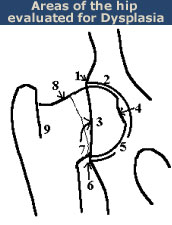
- Cranial acetabular margin
- Femoral head (hip ball)
- Fovea capitus (normal flattened area on hip ball)
- Acetabular notch
- Caudal acetabular rim
- Dorsal acetabular margin
- Junction of femoral head and neck
- Trochanteric fossa
The radiologist is concerned with deviations in these structures from the breed normal. Congruency and confluence of the hip joint (degree of fit) are also considered which dictate the conformation differences within normal when there is an absence of radiographic findings consistent with HD. The radiologist will grade the hips with one of seven different physical (phenotypic) hip conformations: normal which includes excellent, good, or fair classifications, borderline or dysplastic which includes mild, moderate, or severe classifications.
Seven classifications are needed in order to establish heritability information (indexes) for a given breed of dog. Definition of these phenotypic classifications are as follows:
- Excellent
- Good
- Fair
- Borderline
- Mild
- Moderate
- Severe
The hip grades of excellent, good and fair are within normal limits and are given OFA numbers. This information is accepted by AKC on dogs with permanent identification and is in the public domain. Radiographs of borderline, mild, moderate and severely dysplastic hip grades are reviewed by the OFA radiologist and a radiographic report is generated documenting the abnormal radiographic findings. Unless the owner has chosen the open database, dysplastic hip grades are closed to public information.